e99 Online Shopping Mall

Geometry.Net - the online learning center Help  
Home  - Science - Microscopy (Books)

  1-20 of 100 | Next 20
A  B  C  D  E  F  G  H  I  J  K  L  M  N  O  P  Q  R  S  T  U  V  W  X  Y  Z  

click price to see details     click image to enlarge     click link to go to the store

$73.97
1. Transmission Electron Microscopy:
$54.96
2. Scanning Electron Microscopy and
 
3. Fundamentals of Light Microscopy
$64.89
4. Electron Microscopy, 2nd Edition
$55.87
5. Physical Principles of Electron
$60.91
6. Plant Microtechnique and Microscopy
$99.59
7. Introduction to Scanning Tunneling
$85.00
8. Handbook of Biological Confocal
$99.60
9. Scanning Electron Microscopy:
 
$159.00
10. Video Microscopy : The Fundamentals
$96.41
11. Electron Microscopy: Methods and
$55.57
12. Confocal Laser Scanning Microscopy
$51.90
13. Scanning and Transmission Electron
$148.52
14. Optical Imaging and Microscopy:
$58.38
15. Scanning Probe Microscopy:The
 
16. Principles and Techniques of Electron
$27.00
17. Dictionary of Microscopy
 
18. Tissues and Organs: A Text-Atlas
$4.15
19. Scanning Electron Microscopy of
$165.64
20. Raman Microscopy: Developments

1. Transmission Electron Microscopy: A Textbook for Materials Science (4-Vol Set)
by David B. Williams, C. Barry Carter
Paperback: 703 Pages (2004-08-31)
list price: US$95.00 -- used & new: US$73.97
(price subject to change: see help)
Asin: 030645324X
Average Customer Review: 5.0 out of 5 stars
Canada | United Kingdom | Germany | France | Japan
Editorial Review

Book Description
This groundbreaking text provides the necessary instructionsfor hands-on application of this versatile materials characterizationtechnique and is supported by over 600 illustrations and diagrams. ... Read more

Customer Reviews (4)

4-0 out of 5 stars Top of the class
This 4-volume set of paperback books provides a thorough and readable introduction to the science and practice of TEM (transmission electron microscopy).The text is divided into short, digestable sections, each accompanied by figures, graphs and plots.The equations are numerous, but well explained and presented with minimal derivations but full explanations.The books are divided into concise sections making it easy for the reader to find what he/she needs.Overall, a perfect textbook to learn about TEM, and as a reference for those more experienced in this field.

5-0 out of 5 stars Seems like the best TEM book
Good for both beginners and advanced users. Easy to read and well organized. It looks like there are some mistakes in some equations but it is the best TEM book available.

5-0 out of 5 stars Excellent introduction to TEM
In the 70's and 80's the book by Hirsch et al. was the TEM reference tome, and Eddington's book the applications manual.

Time has marched on, and this book is the new replacement for both!

Carter and Williams wrote a very easy to read, yet well detailed, text and reference for TEM.They cover quite literally everything, in just the right level of detail for 1st or 2nd year grad students.

This book is the best way to get a quick grasp of TEM.

5-0 out of 5 stars An outsatnding textbook!
I find this book probably the best textbook in materials science I ever read. Not only the authors are experts in the field of microscopy, but they also succeeded in extremely difficult task to present the complicatedscience of electron microscopy in a simple (but not oversimplified!)language. The book covers all major aspects of transmission electronmicroscopy, and contains excellent illustrations. This book is certainly apriceless asset for students. It is recommended as one of thebest books at the beginning and intermediate level. If you are looking fora book to learn TEM, get this one, you will never regret! ... Read more


2. Scanning Electron Microscopy and X-ray Microanalysis
by Joseph Goldstein, Dale E. Newbury, David C. Joy, Charles E. Lyman, Patrick Echlin, Eric Lifshin, L.C. Sawyer, J.R. Michael
Hardcover: 689 Pages (2003-02)
list price: US$75.00 -- used & new: US$54.96
(price subject to change: see help)
Asin: 0306472929
Average Customer Review: 5.0 out of 5 stars
Canada | United Kingdom | Germany | France | Japan
Editorial Review

Book Description

Providing a comprehensive introduction to the capabilities and use of scanning electron microscopes (SEM) and x-ray spectrometers, this highly acclaimed text emphasizes practical aspects of imaging and analysis for a broad audience of students and practitioners whose backgrounds span a wide range of science and technology. Topics discussed include user-controlled functions of scanning electron microscopes and x-ray spectrometers, the characteristics of electron beam - specimen interactions, image formation and interpretation, the use of x-rays for qualitative and quantitative analysis and the methodology for structural analysis using electron back-scatter diffraction. SEM sample preparation methods for hard materials, polymers, and biological specimens are covered in separate chapters. In addition, techniques for the elimination of charging in non-conducting specimens are detailed. A database of useful parameters for SEM and X-ray micro-analysis calculations and enhancements to the text chapters are available on an accompanying CD.

This third edition has been extensively revised, including new sections on:

  • Variable-pressure SEM,
  • Electron backscatter diffraction (EBSD),
  • Recent developments in x-ray detectors,

and expanded coverage of:

  • Low-voltage SEM,
  • X-ray mapping,
  • Specimen preparation.

The text has been used in educating over 3,000 students at the Lehigh Microscopy School SEM short course as well as thousands of undergraduate and graduate students at universities worldwide.

... Read more

Customer Reviews (6)

4-0 out of 5 stars Excellent outline of SEM and X-Ray microanalysis
This book is a comprehensible review of principles and methods of SEM and X-ray microanalysis write in a single and elegant language. The authors avoid using mathematical formulas in the description and demonstration which turn it an atractive book to all scientists and even the beginners.

5-0 out of 5 stars Great Book !!
This ought to be the dream book of those who do SEM imaging. The first half or say first five/six chapters are solely devoted to fundamentals of SEMs and the rest of the chapters are dealing generation of X-rays and concentrate on EDS. I have not yet finished reading this book. But certainly recommend to other SEM users to possess this book and read it as and when required.

5-0 out of 5 stars Book
The book is very good. I can learn a lot about the SEM from this book. The cd has also some interesting pictures, additional information.

5-0 out of 5 stars The bibel for EM and X-ray Analysis
This book is a great book for learning the Basics about Electron Microscopy and X-ray Analysis. You get a good overview!

5-0 out of 5 stars Scanning Electron Microscopy Book
The book came in excellent condition as stated. It also arrived in a timely manner. ... Read more


3. Fundamentals of Light Microscopy and Electronic Imaging
by Douglas B. Murphy
 Hardcover: 432 Pages (2008-04-11)

Isbn: 047169214X
Average Customer Review: 5.0 out of 5 stars
Canada | United Kingdom | Germany | France | Japan
Editorial Review

Book Description
Over the last decade, advances in science and technology have profoundly changed the face of light microscopy. Research scientists need to learn new skills in order to use a modern research microscope–skills such as how to align microscope optics and perform image processing. Fundamentals of Light Microscopy and Electronic Imaging explores the basics of microscope design and use. The comprehensive material discusses the optical principles involved in diffraction and image formation in the light microscope, the basic modes of light microscopy, the components of modern electronic imaging systems, and the image processing operations necessary to acquire and prepare an image.

Written in a practical, accessible style, Fundamentals of Light Microscopy and Electronic Imaging reviews such topics as:

  • Illuminators, filters, and isolation of specific wavelengths
  • Phase contrast and differential interference contrast
  • Properties of polarized light and polarization microscopy
  • Fluorescence and confocal laser scanning microscopy
  • Digital CCD microscopy and image processing

Each chapter includes practical demonstrations and exercises along with a discussion of the relevant material. In addition, a thorough glossary assists with complex terminology and an appendix contains lists of materials, procedures for specimen preparation, and answers to questions.

An essential resource for both, experienced and novice microscopists.Download Description
This practical book presents the fundamentals of light microscopy, including the basics of microscope design, image formation, and camera function. Moreover, researchers will learn how to acquire electronic images and perform image processing. The author provides instructions on the latest techniques involving confocal laser scanning microscopy, digital CCD microscopy, as well as a chapter covering issues related to image processing for scientific publication. ... Read more

Customer Reviews (4)

4-0 out of 5 stars A little dated, but a good textbook
I had to buy this book for a Graduate level course that I took.We covered all of the material in the book in about 4 weeks.I didn't know anything about the class beforehand, but the book did help a lot. It was a little dull, but the content was good albeit a little outdated.

5-0 out of 5 stars Keep this book by the microscope
This is the book that tells you how to actually do effective microscopy.Keep a copy by the microscope.

5-0 out of 5 stars Best Introductory-Level Textbook
Dr. Murphy constructs a solid foundation on the basic concepts of geometrical optics, light, and color, and then provides excellent introductory reviews of important topics in light microscopy.The book is very well written and complex phenomena are clearly explained without the unnecessary math that often confuses students.Illustrations are numerous and help support the text very nicely, as do the suggested laboratory exercises that accompany each chapter.Discussions of digital cameras and image processing are timely and provide the essential concepts necessary to tackle more advanced treatises.In the opinion of the Molecular Expressions microscopy website team, this book is by far the best entry-level textbook in the field.

5-0 out of 5 stars Well Written and Useful
"This well written text provides a clear, uncluttered overview of the principles and practice of modern light microscopy. It contains many helpful teaching exercises and diagrams. It should prove useful in a wide range of courses from the undergraduate to the postgraduate level."
-- Kenneth R. Spring, Ph.D., Author ... Read more


4. Electron Microscopy, 2nd Edition
by John J. Bozzola, Lonnie Dee Russell
Hardcover: 670 Pages (1998-10)
list price: US$119.95 -- used & new: US$64.89
(price subject to change: see help)
Asin: 0763701920
Average Customer Review: 5.0 out of 5 stars
Canada | United Kingdom | Germany | France | Japan
Editorial Review

Book Description
Electron Microscopy covers all of the important aspects of electron microscopy for biologists, including theory of scanning and transmission, specimen preparation, digital imaging and image analysis, laboratory safety and interpretation of images. The text also contains a complete atlas of ultrastructure. ... Read more

Customer Reviews (4)

4-0 out of 5 stars Electron microscopy for biologists
This book is a very good technical introduction to the electron microscopy of biological samples. Good figures and schemes help to understand the text. Photos of different types of samples are also adequate to illustrate the methods. The book is an excellent manual for beginners and biologists find it useful and easy to read.

5-0 out of 5 stars Excellent Book
This book is excellent for beginners and advanced microscopists. It offers fine details and tricks about techniques as well as the theory behind it. There are lots of pictures and schematics for each descriptions. It is also useful for microscopist that want to learn more about different methods. I definitively recommend it!

5-0 out of 5 stars Excellent EM primer
I actually had the opportunity to receive instruction from Prof. Bozzola in both Transmission Electron Microscopy (TEM) and Scanning Electron Microscopy (SEM), and acquired the book for those reasons.It is not common to find even a textbook with such well-detailed instructions from beginning level to advanced for a myriad of topics in the field.Of course, there is a very significant focus on biological applications, so some portions of the book are not entirely relevant to materials science folks like myself.This book covers the basics of optics, sample preparation, imaging, photography and film developing, and specifics about both TEMs and SEMs.This book is a very good investment for anyone intending to perform work with such instruments.

5-0 out of 5 stars If you are learning EM you must have this book...
This is a great book for those entering the field of EM study. It takes you step by step through both SEM and TEM sample prep and analysis. It is packed with helpful pictures and diagrams. It will be a book you will referto for methodology and protocal from fixation to sectioning...I really lovethis book. ... Read more


5. Physical Principles of Electron Microscopy: An Introduction to TEM, SEM, and AEM
by Ray F. Egerton
Hardcover: 202 Pages (2008-05-15)
list price: US$69.95 -- used & new: US$55.87
(price subject to change: see help)
Asin: 0387258000
Canada | United Kingdom | Germany | France | Japan
Editorial Review

Book Description

Scanning and stationary-beam electron microscopes have become an indispensable tool for both research and routine evaluation in materials science, the semiconductor industry, nanotechnology, and the biological and medical sciences. Physical Principles of Electron Microscopy provides an introduction to the theory and current practice of electron microscopy for undergraduate students who want to acquire an appreciation of how basic principles of physics are utilized in an important area of applied science, and for graduate students and technologists who make use of electron microscopes. At the same time, this book will be equally valuable for university teachers and researchers who need a concise supplemental text that deals with the basic principles of microscopy.

... Read more

6. Plant Microtechnique and Microscopy
by Steven E. Ruzin
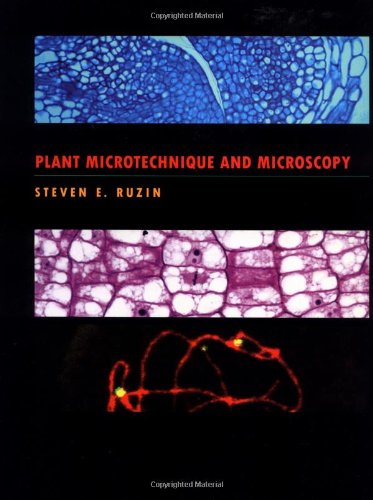
Paperback: 336 Pages (1999-05-20)
list price: US$67.95 -- used & new: US$60.91
(price subject to change: see help)
Asin: 0195089561
Average Customer Review: 5.0 out of 5 stars
Canada | United Kingdom | Germany | France | Japan
Editorial Review

Book Description
Plant microtechnique has generated renewed interest in recent years, due in part to the need for molecular biologists to visualize a gene or gene product in the context of the whole plant. Plant Microtechnique and Microscopy offers uniquely in-depth coverage of this reinvigorated field. Thoroughly covering classical aspects of microscope slide preparation, it goes a step beyond all other available manuals by also documenting the theory and practice of modern applications.The text opens with single-page "Quick Start" protocols that provide students with fundamental instructions to complete eight of the most common microtechnique protocols used today. The following sections cover the theory and practice of microtechnique. The traditional paraffin method is demonstrated by explicit step-by-step protocols, and theoretical background is incorporated to give students the tools required to design their own experiments and to interpret existing results. In addition, modern applications such as methacrylate embedding and sectioning, microwave tissue processing, fluorescence histochemistry, and in situ hybridization are discussed in detail. The manual also contains a definitive chapter on microscopy and describes, in both text and diagrams, the optical principles of techniques such as phase contrast and DIC as well as confocal and deconvolution wide-field microscopy. Appendices on laboratory practice (chemical toxicities, common calculations, and buffer tables), an extensive appendix on optics and its application to the microscope, and an extensive bibliography of over 550 references are also included. Ideal for courses in plant biology, Plant Microtechnique and Microscopy also serves as an indispensable reference for all students of microscopy, histology, and histological technique. It is a valuable addition to every biological laboratory. ... Read more

Customer Reviews (1)

5-0 out of 5 stars NEW STANDARD FOR PLANT MICROTECHNIQUE
I loved it. IT WAS MUCH BETTER THAN CATS... I'M GOING TO READ IT AGAIN AND AGAIN...... ... Read more


7. Introduction to Scanning Tunneling Microscopy (Monographs on the Physics and Chemistry of Materials)
by C. Julian Chen
Hardcover: 432 Pages (2007-12-05)
list price: US$130.00 -- used & new: US$99.59
(price subject to change: see help)
Asin: 0199211507
Average Customer Review: 5.0 out of 5 stars
Canada | United Kingdom | Germany | France | Japan
Editorial Review

Book Description
The scanning tunneling microscope and the atomic force microscope, both capable of imaging and manipulating individual atoms, were crowned with the Nobel Prize in Physics in 1986, and are the cornerstones of nanotechnology today. The first edition of this book has nurtured numerous beginners and experts since 1993. The second edition is a thoroughly updated version of this 'bible' in the field. The second edition includes a number of new developments in the field. Non-contact atomic-force microscopy has demonstrated true atomic resolution. It enables direct observation and mapping of individual chemical bonds. A new chapter about the underlying physics, atomic forces, is added. The chapter on atomic force microscopy is substantially expanded. Spin-polarized STM has enabled the observation of local magnetic phenomena down to atomic scale. A pedagogical presentation of the basic concepts is included. Inelastic scanning tunneling microscopy has shown the capability of studying vibrational modes of individual molecules. The underlying theory and new instrumentation are added. For biological research, to increase the speed of scanning to observe life phenomena in real time is a key. Advanced in this direction is presented as well. The capability of STM to manipulate individual atoms is one of the cornerstones of nanotechnology. The theoretical basis and in particular the relation between tunneling and interaction energy are thoroughly presented, together with experimental facts. ... Read more

Customer Reviews (1)

5-0 out of 5 stars Great resource for amateur STM designers
This may be a good resource for professionals also - but they'll have to speak for themselves. As an amateur, I found the instrumentation section extremely valuable. It is primarily because of that section that I give the text 5 stars. After plowing through a great many journal papers I eventually found that most of the theory and practice of building an STM had in fact been nicely distilled by Chen. There are some minor mistakes (e.g. discrepencies between the text and some figures), but nothing critical that I could see. A number of disparate but relevant prerequisite topics are addressed in the appendices with moderate success.
If there is a peer or superior alternative to the instrumentation and design aspects of STMs, I would love to hear of it! ... Read more


8. Handbook of Biological Confocal Microscopy
Hardcover: 988 Pages (2006-06-02)
list price: US$129.00 -- used & new: US$85.00
(price subject to change: see help)
Asin: 038725921X
Average Customer Review: 5.0 out of 5 stars
Canada | United Kingdom | Germany | France | Japan
Editorial Review

Book Description

This third edition of a classic text in biological microscopy includes detailed descriptions and in-depth comparisons of parts of the microscope itself, digital aspects of data acquisition and properties of fluorescent dyes, the techniques of 3D specimen preparation and the fundamental limitations, and practical complexities of quantitative confocal fluorescence imaging.

... Read more

Customer Reviews (2)

5-0 out of 5 stars A Collection Of Great Papers
This is a great Handbook that contains many well written papers on various aspects of Confocal Microscopy, with emphasis on biological samples.There are basic and tutorial articles as well as some detailed specific studies.Many color photographs and charts are also included where appropriate.I highly recommend it to novice and expert workers in this field.For the former, it will provide a decent foundation, especially with references that are in each article; and for the latter, it serves as a compendium of good practices.

5-0 out of 5 stars A must have if you want to know about confocal microscopy
This is a great reference book.A great place to start if you want to learn about confocal microscopes.The chapters are written by experts in their specific field of confocal microscopy. The book is also wellreferenced so that you can easliy continue your search for knowledge onconfocal microscopy.I also found the appendix interesting, they list mostof the existing confocal systems and there specs.This is useful if youplan on purchasing one.This book is also useful if you plan on buildingyour own. ... Read more


9. Scanning Electron Microscopy: Physics of Image Formation and Microanalysis (Springer Series in Optical Sciences)
by Ludwig Reimer
Hardcover: 527 Pages (1998-10-16)
list price: US$125.00 -- used & new: US$99.60
(price subject to change: see help)
Asin: 3540639764
Canada | United Kingdom | Germany | France | Japan
Editorial Review

Book Description
Scanning Electron Microscopy provides a description of the physics of electron-probe formation and of electron-specimen interactions. The different imaging and analytical modes using secondary and backscattered electrons, electron-beam-induced currents, X-ray and Auger electrons, electron channelling effects, and cathodoluminescence are discussed to evaluate specific contrasts and to obtain quantitative information. ... Read more


10. Video Microscopy : The Fundamentals (The Language of Science)
by Shinya Inoué, Kenneth R. Spring
 Hardcover: 770 Pages (1997-08-31)
list price: US$159.00 -- used & new: US$159.00
(price subject to change: see help)
Asin: 0306455315
Average Customer Review: 4.5 out of 5 stars
Canada | United Kingdom | Germany | France | Japan
Editorial Review

Book Description
This classic has been throughly revised and expanded to reflecttechnological advances, but it still emphasizes the fundamentals, withextensive reviews of optical microscopy, video detectors, and displaydevices. Three completely new chapters deal with solid-statedetectors, both video-rate and slow scan, image intensifiction; andthe color video signal and color video cameras. Another new chaptertreating the practical aspcts of the design and use of videomicroscope systems is especially notable. This edition fetures over500 illustrations (including 126 half-tones), a glossary of items,detailed tables of specifications, and a list of manufacturers. ... Read more

Customer Reviews (3)

4-0 out of 5 stars video microscopy review
Book was informative for basics and advanced material.
I found it difficult to read; was too much info for a basic microscopy course. I think there should be an easier to read edition that focuses more on microscopy techniques without all the advanced information.

5-0 out of 5 stars An invaluable reference
The author provides a very detailed and extremely well presented guide to all aspects of video microscopy. The subject is presented in such a way as to be of great value to the most advanced reader while not being beyond the grasp of the novice. All relevant background material is supplied, and I have found the book to be an excellent reference for video imaging and image processing in general.

5-0 out of 5 stars EXCELLENT BOOK!!
This book is an excellent update to the first edition.Much progress has been made in Video Microscopy since the first edition in 1986.This book covers the field nicely.It is an excellent guide for the novice and excellent reference for the advanced user.Anyone serious about video microscopy should have a copy of this book on their shelf! ... Read more


11. Electron Microscopy: Methods and Protocols (Methods in Molecular Biology)
Hardcover: 608 Pages (2007-02-27)
list price: US$150.00 -- used & new: US$96.41
(price subject to change: see help)
Asin: 1588295737
Average Customer Review: 4.0 out of 5 stars
Canada | United Kingdom | Germany | France | Japan
Editorial Review

Book Description
In this revised and expanded second edition, Electron Microscopy: Methods and Protocols presents the newest technology in electron microscopy, while maintaining the practicality and accessibility of the acclaimed first edition.Like the first edition, this volume provides clear, concise instructions on processing biological specimens and includes discussion on the underlying principles of the majority of the processes presented.Each protocol is presented by experts with years of first-hand experience and in-depth knowledge of the techniques they describe.The Notes section accompanying each protocol will prove invaluable for adapting and troubleshooting protocols.

Electron Microscopy: Methods and Protocols, Second Edition, comprises two major areas of electron microscopy-transmission electron microscopy (TEM) and scanning electron microscopy (SEM).The TEM area covers several key techniques, including: conventional specimen preparation methods for cultured cells and biomedical and plant tissues; cryospecimen preparation by high-pressure freezing and cryoultramicrotomy negative staining and immunogold labeling techniques; and TEM crystallography and cryo-TEM tomography.The SEM area similarly attends to conventional-, variable pressure-, environmental-, and cryoscanning microscopy techniques, as well as the application of X-ray microanalysis.Protocols for the application of X-ray microanalysis to SEM and mass spectrometry conclude the volume.

... Read more

Customer Reviews (1)

4-0 out of 5 stars A good lab reference book
This book covers pretty much all of the modern techniques used in biological electron microscopy. It emphasizes the availability of many protocols for both Transmission and Scanning EM. Every chapter has a detailed protocol anda full list of accompanying references, in case the reader wants to know more about a specific topic/technique.

It is highly recommended for every student, lab tech, or researcher that wants to have an overview of the existing modern techniques or dare trying a new technique. It's format, with its 600+ pages is not very practical as a lab handbook, but the content information of strict high quality.

I have more than 10yrs of experience in biological cryo-EM, and I find this book a very useful addition to my ever growing collection. ... Read more


12. Confocal Laser Scanning Microscopy (Microscopy Handbooks)
by C. J. R. Sheppard, D. M. Hotton, David Shotton
Paperback: 106 Pages (1997-08-31)
list price: US$52.95 -- used & new: US$55.57
(price subject to change: see help)
Asin: B000HWYY8K
Average Customer Review: 3.5 out of 5 stars
Canada | United Kingdom | Germany | France | Japan

Customer Reviews (3)

4-0 out of 5 stars Good intro to the instrument
I don't need to use the microscope, I just need to understand the instrument and the 3D images it produces. This book gave me the introduction I wanted.

It starts with a brief discussion the basics: illumination, scanning, and rejecting out-of-plane light. The next chapter describes a bit more about the pieces that go into the instrument and a little about the optical theory. That chapter is nowhere near what an instrument designer would need to know. It does, however, give a user enough information to use the tool effectively. The next sections discuss fluorescence, including the problems that arise when handling long and short wavelengths at through the same optics, and a little about specimen prepration using nanoparticles. The final sections briefly discuss applications and possibilities for the future of confocal microscopy.

The authors do not have much to say about specific protocols for specific applications - the level is more general and centered more on the microscope itself. Anyone who wants a how-to book for the lab bench is going to be disappointed. I wanted different information, and I got what I wanted from it.

//wiredweird

2-0 out of 5 stars Don't use this for Biological Applications
I was dissappointed by this book.It has very little about applications, and more about the physics of the microscope. I find better explanations of the basic concept of CLSM at web sites of some of the microscope manufacturers like [manufacture names]...

4-0 out of 5 stars CLSM for Dummies
An excellent primer for those who have just a little knowledge, but are missing the background to handle CLSM data with confidence.Readers with some knowledge of the jargon of microscopy will find the book to be aneasier read, but this reviewer had no prior knowledge and found the bookextremely helpful.It gives enough fundamental information to let thepotential CLSM user know where s/he is going to get into trouble.If youare going to image something for the first time, get this book.It'll keepyou from interpreting artifacts. ... Read more


13. Scanning and Transmission Electron Microscopy: An Introduction
by Stanley L. Flegler, John W. Heckman, Karen L. Klomparens
Hardcover: 240 Pages (1993-09-23)
list price: US$67.95 -- used & new: US$51.90
(price subject to change: see help)
Asin: 0195107519
Average Customer Review: 5.0 out of 5 stars
Canada | United Kingdom | Germany | France | Japan
Editorial Review

Book Description
This authoritative volume, ideal for use in the laboratory, presents the practical and theoretical fundamentals of scanning and transmission electron microscopy--together in one convenient volume. Clear and concise explanations coupled with instructive diagrams and photographs guide you through microscope operation, image production, analytical techniques, and potential applications to various disciplines.Specimen preparation is discussed in detail, with emphasis on specific parameters for biological specimens.Since each laboratory has its own procedures, this unique book covers the essentials of scanning and transmission electron microscopy while leaving the laboratory particulars to individual discretion.Unmatched in scope and clarity--and filled with helpful diagrams, photographs, and drawings--this text offers the best introduction to scanning and transmission electron microscopy available.Due to its comprehensive coverage, the book will serve as an ideal course text in the electron microscopy classes organized for the benefit of advanced students in both the biological and physical sciences. ... Read more

Customer Reviews (1)

5-0 out of 5 stars A great intro to SEM/TEM!
As an SEM/EDS (read the book) process engineer working with technicians at many different levels, I have found this book to be an invaluable resource.It is written at very basic level making it accessible to anyone, with or without a scientific background.Whenever someone wants to know more about how the SEM functions then I can tell them in a five minute spiel, I give them a copy of this book.The material is slightly dated (early 90's technology), but is still relevant to anyone in the field today.A great book on all of the basics. ... Read more


14. Optical Imaging and Microscopy: Techniques and Advanced Systems (Springer Series in Optical Sciences) (Springer Series in Optical Sciences)
Hardcover: 500 Pages (2007-09-14)
list price: US$189.00 -- used & new: US$148.52
(price subject to change: see help)
Asin: 354069563X
Canada | United Kingdom | Germany | France | Japan
Editorial Review

Product Description

This text on contemporary optical systems is intended for optical researchers and engineers, graduate students and optical microscopists in the biological and biomedical sciences. This second edition contains two completely new chapters. In addition most of the chapters from the first edition have been revised and updated. The book consists of three parts: The first discusses high-aperture optical systems, which form the backbone of optical microscopes. An example is a chapter new in the second edition on the emerging field of high numerical aperture diffractive lenses which seems to have particular promise in improving the correction of lenses. In this part particular attention is paid to optical data storage. The second part is on the use of non-linear optical techniques, including nonlinear optical excitation (total internal reflection fluorescence, second and third harmonic generation and two photon microscopy) and non-linear spectroscopy (CARS). The final part of the book presents miscellaneous techniques that are either novel or well known but finding new applications. An example of the latter is photonic force microscopy that is now rapidly becoming an exciting and indispensable tool in shedding light on the inner working of certain cells or measuring nano-mechanical properties of single molecules.

... Read more

15. Scanning Probe Microscopy:The Lab on a Tip
by Ernst Meyer, Hans J. Hug, Roland Bennewitz
Hardcover: 210 Pages (2003-10-10)
list price: US$79.95 -- used & new: US$58.38
(price subject to change: see help)
Asin: 3540431802
Average Customer Review: 4.0 out of 5 stars
Canada | United Kingdom | Germany | France | Japan
Editorial Review

Book Description

Written by three leading experts in the field, this textbook describes and explains all aspects of the scanning probe microscopy. Emphasis is placed on the experimental design and procedures required to optimize the performance of the various methods. Scanning Probe Microscopy covers not only the physical principles behind scanning probe microscopy but also questions of instrumental designs, basic features of the different imaging modes, and recurring artifacts. The intention is to provide a general textbook for all types of classes that address scanning probe microscopy. Third year undergraduates and beyond should be able to use it for self-study or as textbook to accompany a course on probe microscopy. Furthermore, it will be valuable as reference book in any scanning probe microscopy laboratory. Novel applications and the latest important results are also presented, and the book closes with a look at the future prospects of scanning probe microscopy, also discussing related techniques in nanoscience. Ideally suited as an introduction for graduate students, the book will also serve as a valuable reference for practising researchers developing and using scanning probe techniques.

... Read more

Customer Reviews (1)

4-0 out of 5 stars Solid Background to SPM
Scanning Probe Microscopy: The Lab on a Tip by Ernst Meyer covers the two main techniques of SPM: STM and AFM (here called SFM). There is also a detailed section on Magnetic Force Microscopy(MFM). There are shorther sections on the less well known techniques, i.e. friction force microsocpy, SNOM, etc. The books covers the theory well, and is reasonably up to date as I write in 2007. The figures are clear, and the few microgrpahs presented in clear greyscale. There are relatively few applications, but these are easy to find elsewhere. Overall, a great book for the theory of SPM. ... Read more


16. Principles and Techniques of Electron Microscopy: Biological Applications
by M. A. Hayat
 Hardcover: Pages (1990-01)
list price: US$71.00
Isbn: 0849371112
Average Customer Review: 5.0 out of 5 stars
Canada | United Kingdom | Germany | France | Japan
Editorial Review

Book Description
Principles and Techniques of Electron Microscopy is the standard work for biological electron microscopists wishing to learn how to prepare their specimens for electron microscopic investigation.This fully revised and expanded fourth edition includes three new chapters covering such topics as plant tissues, immunocytochemistry, and applications of microwave irradiation to microscopy. It provides practical instructions on how to process biological specimens, as well as a detailed discussion on the principles underlying the various processes.Dr. Hayat presents methods in a self-explanatory form and includes alternative procedures and points of disagreement to help the reader interpret data accurately.What sets this book apart from its competition is that it not only describes techniques but also explains their fundamental principles; that is, those chemical reactions underlying the use of various reagents for preserving and staining cellular components. ... Read more

Customer Reviews (1)

5-0 out of 5 stars labeling with colloidal gold conjugates
It is an excellent handbook for electron microscopy. In the section of labeling with protein A / colloidal gold, the author fails to acknowledge the original contribution on the subject: Romano EL and Romano MS: Staphylococcal protein A bound to colloidal gold: a useful reagent to label antigen-antibody sites in electron microscopy. Immunochemistry 14:711-715,1977. I think that the omission is unfair because later works by others are cited in the same section of the book ... Read more


17. Dictionary of Microscopy
by Julian P. Heath
Paperback: 358 Pages (2005-09-30)
list price: US$60.00 -- used & new: US$27.00
(price subject to change: see help)
Asin: 0470011998
Canada | United Kingdom | Germany | France | Japan
Editorial Review

Book Description
The past decade has seen huge advances in the application of microscopy in all areas of science. This welcome development in microscopy has been paralleled by an expansion of the vocabulary of technical terms used in microscopy: terms have been coined for new instruments and techniques and, as microscopes reach even higher resolution, the use of terms that relate to the optical and physical principles underpinning microscopy is now commonplace.

The Dictionary of Microscopy was compiled to meet this challenge and provides concise definitions of over 2,500 terms used in the fields of light microscopy, electron microscopy, scanning probe microscopy, x-ray microscopy and related techniques. Written by Dr Julian P. Heath, Editor of Microscopy and Analysis (http://www.microscopy-analysis.com),  the dictionary is intended to provide easy navigation through the microscopy terminology and to be a first point of reference for definitions of new and established terms.

The Dictionary of Microscopy is an essential, accessible resource for:

  • students who are new to the field and are learning about microscopes
  • equipment purchasers who want an explanation of the terms used in manufacturers' literature
  • scientists who are considering using a new microscopical technique
  • experienced microscopists as an aide mémoire or quick source of reference
  • librarians, the press and marketing personnel who require definitions for technical reports.
... Read more

18. Tissues and Organs: A Text-Atlas of Scanning Electron Microscopy
by Richard G. Kessel
 Paperback: 317 Pages (1979-02)
list price: US$54.85
Isbn: 0716700905
Average Customer Review: 5.0 out of 5 stars
Canada | United Kingdom | Germany | France | Japan

Customer Reviews (2)

5-0 out of 5 stars This is a must book for Histology
I really enjoyed the book. It goes very well with Histology courses.It gives you feeling of three dimensional pictures of real cells like photography. Since it is like a photography, it shows external views not the section throughs but you get hundered times better understanding of the structures. just look at scanning electron picture examples in your typical cell books. This book is full of them and not just plain pictures but with some explanations similar to Histology books.

5-0 out of 5 stars an indispensable reference
This is a fantastic visual reference book for anybody interested in microscopic and/or macroscopic anatomy. Most of the major systems are covered, although the tissue samples are usually taken from small mammals.The range of magnification is great, and provides the reader with a clearconcept of the structure and function of a given organ.

I'm aprofessional medical illustrator with a library of over 300 books, and thisis one of only a few "desert island" books - ones that I wouldn'twant to be without. I believe anybody with an interest in the way we'remade will find it fascinating. ... Read more


19. Scanning Electron Microscopy of Cerebellar Cortex
by Orlando Castejón
Paperback: 148 Pages (2003-06-30)
list price: US$113.00 -- used & new: US$4.15
(price subject to change: see help)
Asin: 0306477114
Canada | United Kingdom | Germany | France | Japan
Editorial Review

Book Description

In a clear exposition this inspirational book shows the in situ three-dimensional morphology of cerebellar neurons, intracortical circuits and synaptic connections that underpin the functioning of cerebellar neurons in networks. It brings together in one volume a new view of the three-dimensional cytoarchitectonic arrangement of the cerebellar cortex.

The book shows the cerebellar extrinsic and intrinsic intracortical circuits formed by mossy and climbing fibers as exposed by the cryofracture methods. The high degree of lateral collateralization of these fibers is also displayed providing new insights on the information processing in the cerebellar cortex. Besides, field emission high resolution electron microscopy shows its potential contribution to the study of synaptic morphology. The concluding chapter deals with the contribution of scanning electron microscopy to cerebellar neurobiology.

This monograph is an authoritative survey and a must for anyone who is interested in the structure of the central nervous system. It will also appeal to an interdisciplinary audience who wants to learn more about electron microscopy and neurocytology.

... Read more

20. Raman Microscopy: Developments and Applications
Hardcover: 463 Pages (1996-07-03)
list price: US$205.00 -- used & new: US$165.64
(price subject to change: see help)
Asin: 0121896900
Canada | United Kingdom | Germany | France | Japan
Editorial Review

Book Description
One of the first books devoted entirely to the subject of Raman microscopy, this volume addresses issues of great interest to engineers working in Raman-microscope development and researchers concerned with areas ofapplication for this science. The book is written by several world recognized experts, who summarize the Raman effect before discussing the hardware and software involved in todays instruments. This format provides an excellent introduction to this up-and-coming discipline. All important applications, including those in materials science and earth science are covered in depth.

Includes extensive description of the instrumentation, the Raman microspectrograph, the treatment of data, and micro-Raman imaging
Examines the use of Raman microscopy in diverse applications, including some of the hyphenated methods
Summarizes the Raman effect
Discusses new uses for this technology ... Read more


  1-20 of 100 | Next 20
A  B  C  D  E  F  G  H  I  J  K  L  M  N  O  P  Q  R  S  T  U  V  W  X  Y  Z  

Prices listed on this site are subject to change without notice.
Questions on ordering or shipping? click here for help.

site stats